Specialized Neurodiagnostic & Vascular Evaluations
• Advanced, in-office testing to assess brain wave activity, peripheral nerve function, and cranial blood flow
• Advanced, in-office testing to assess brain wave activity, peripheral nerve function, and cranial blood flow
Electroencephalogram (EEG)
Also known as a Brain Wave Test
Overview
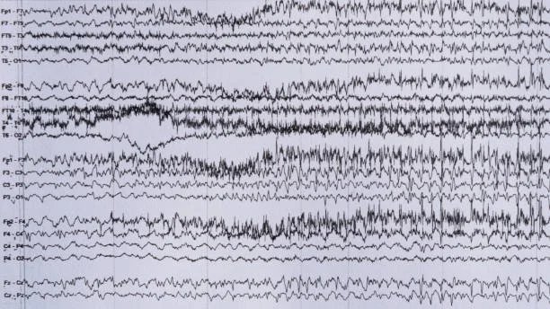
An Electroencephalogram (EEG) is a non-invasive diagnostic test that measures and records the spontaneous electrical activity generated by brain cells (neurons). By analyzing brain wave patterns, physicians can detect abnormalities associated with:
Seizure disorders and epilepsy
Unexplained loss of consciousness, fainting, or altered mental status
Sleep disorders and encephalopathy
Memory decline and structural or metabolic cerebral conditions
How to Prepare
Hair & Scalp Hygiene: Wash your hair thoroughly with shampoo the night before or morning of your test. Do not apply hairspray, gel, oils, leave-in conditioners, or pomades.
Medications: Take your routine prescribed medications unless your physician provides alternative instructions.
Transportation Safety: If you have a history of frequent seizures, episodes of confusion, or are undergoing sleep-deprivation testing, please arrange for a companion or driver to take you to and from the clinic.
What to Expect During the Procedure
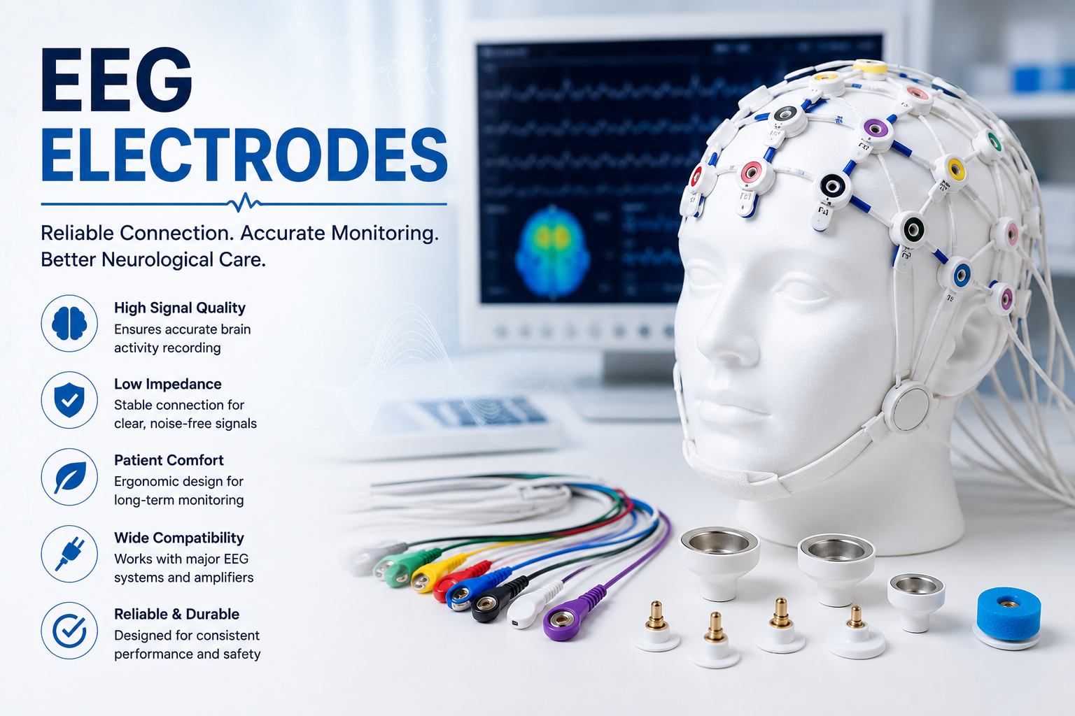
Small, flat sensor discs (electrodes) are attached to specific locations on your scalp using a temporary, water-soluble adhesive paste.
The electrodes detect micro-electrical signals from your brain and transmit them to a high-resolution recording monitor.
The test is completely painless and typically takes 30 to 60 minutes while you relax comfortably in an exam chair.
Transcranial Doppler (TCD)
Cranial Blood Flow Assessment
Overview
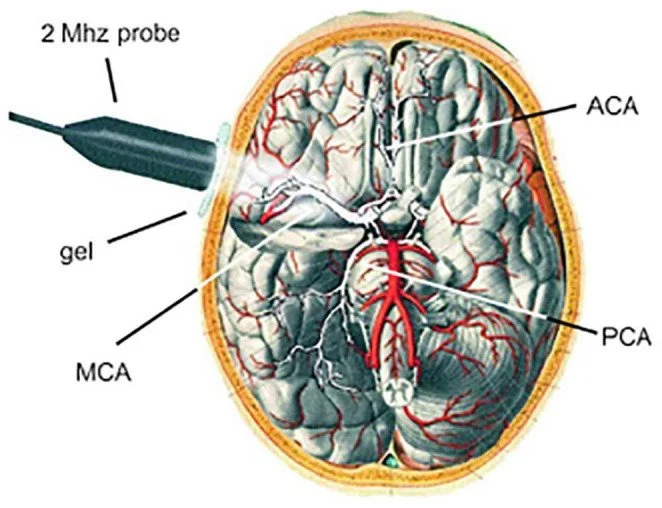
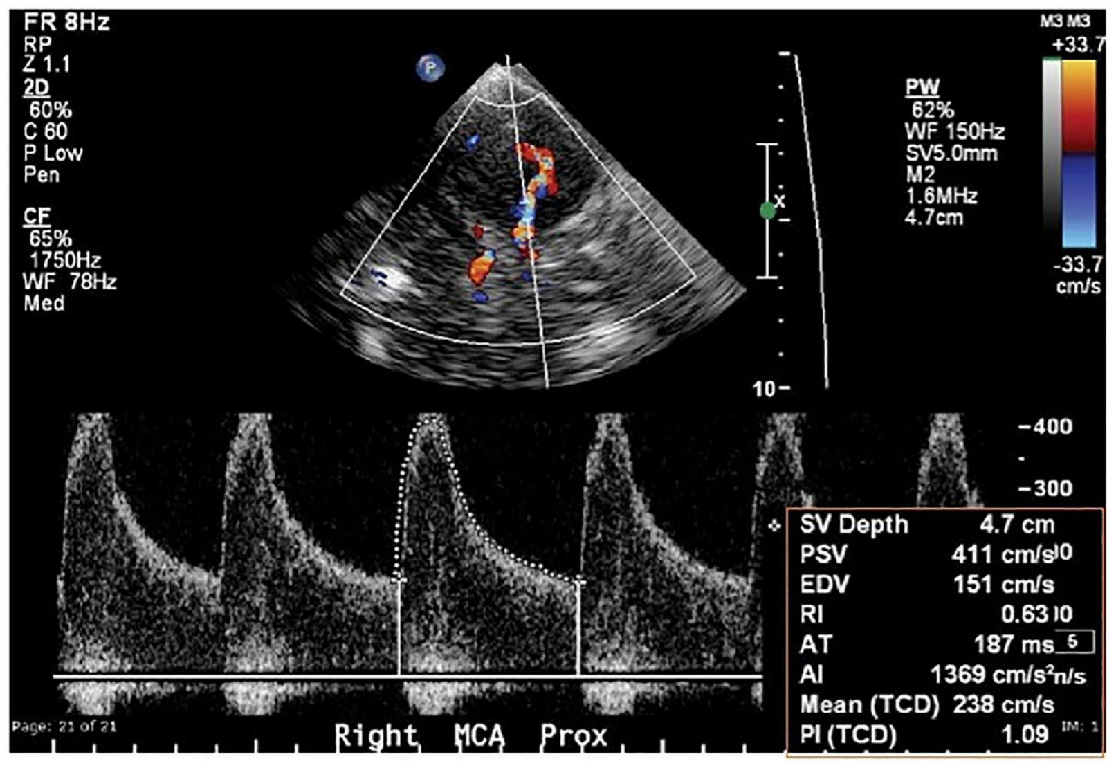
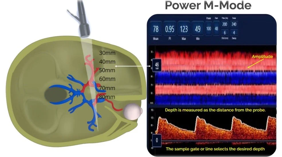
A Transcranial Doppler (TCD) is a painless, specialized ultrasound examination that assesses the velocity, direction, and resistance of blood flow within the intracranial arteries supplying the brain. It is an essential clinical tool used to detect arterial narrowing (stenosis), microemboli, vasospasm, and collateral circulation patterns.
What to Expect During the Procedure
Non-Invasive Acoustic Windows: The technologist applies a water-soluble acoustic gel to specific areas where cranial bone is naturally thinner, allowing ultrasound waves to penetrate clearly:
Temples on either side of the head
The nape / base of the neck
Underneath the chin
Gently over closed eyelids
Exam Duration: A standard TCD evaluation takes approximately 30 to 45 minutes.
Physiologic Compression Maneuvers: In select clinical situations, the technician may briefly apply gentle pressure to a carotid artery in the neck to observe dynamic shifts in intracranial blood flow.
Aftercare
There are no recovery restrictions. The acoustic gel easily washes off with mild soap and warm water, and you may immediately return to regular daily activities.
Nerve & Muscle Assessment: NCS & EMG
Electromyography and Nerve Conduction Studies
Overview
Nerve Conduction Studies (NCS) and Electromyography (EMG) are specialized diagnostic tools evaluated in tandem to assess the health and electrical signaling of your peripheral nerves and muscles. This evaluation pinpoints conditions such as peripheral neuropathy, carpal tunnel syndrome, cervical/lumbar radiculopathy (pinched nerves), and myopathy.
This evaluation is conducted in two complementary parts:
Part 1: Nerve Conduction Study (NCS)
Purpose: Measures the integrity, conduction velocity (speed), and signal amplitude of sensory and motor nerves.
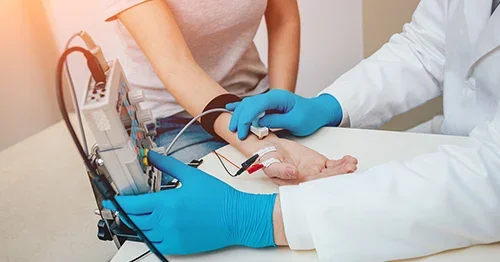
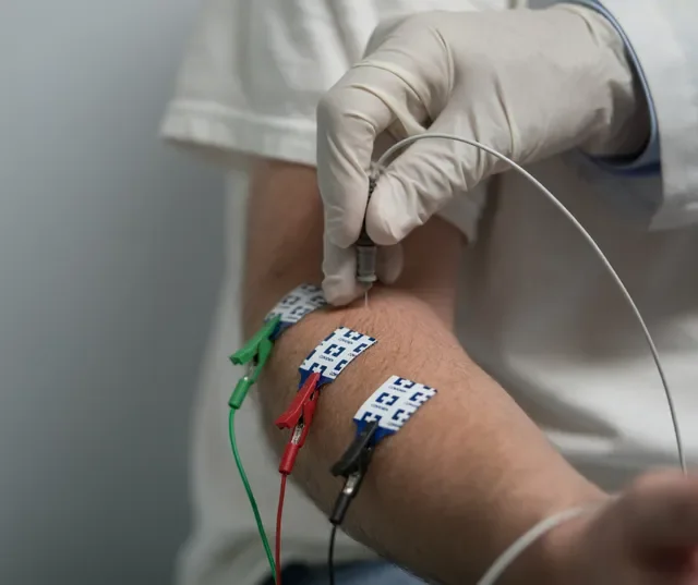
What to Expect: Small adhesive surface electrodes are placed on your arms, hands, legs, or feet. Mild, brief electrical pulses stimulate specific nerves while the sensor electrodes record how quickly and effectively the signal travels along the nerve pathway.
Part 2: Electromyogram (EMG)
Purpose: Analyzes the electrical activity of muscle fibers at rest and during voluntary muscle contraction.
What to Expect: A fine, sterile pin electrode is gently inserted into targeted muscles to evaluate muscle motor units. You may experience a brief sensation comparable to a quick injection. Remaining calm and relaxed during the test ensures minimal discomfort and optimal diagnostic precision.
Carotid Duplex Ultrasound Scan
Extracranial Vascular Assessment
Overview
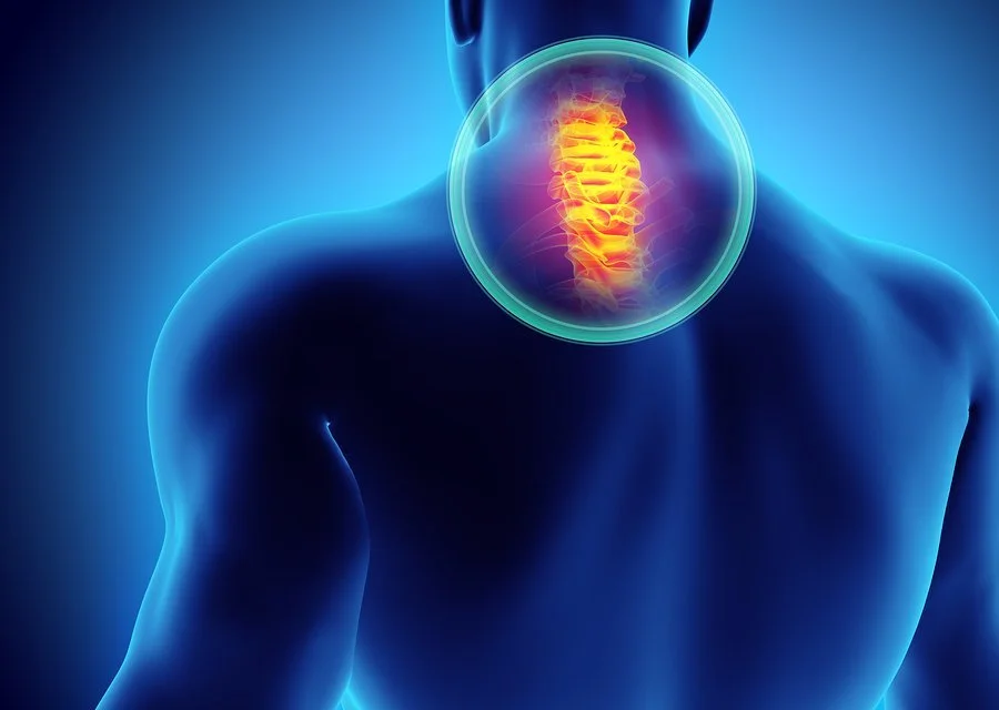
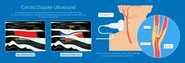
A Carotid Duplex Scan is a painless, high-resolution diagnostic ultrasound that visualizes the carotid and vertebral arteries in the neck. These major vessels supply oxygen-rich blood directly from the heart to the brain.
Clinical Value & Purpose
Detecting Arterial Plaque: Identifies atheromatous plaque accumulation along the arterial wall.
Assessing Stenosis & Occlusion: Measures the degree of narrowing to accurately calculate stroke and TIA risk.
Guiding Intervention: Assists your vascular neurologist in determining whether medical management, endovascular intervention, or surgical referral is indicated.
What to Expect
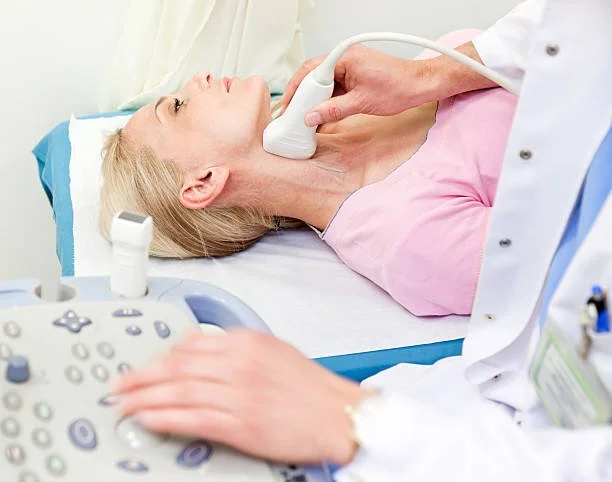
You will lie comfortably on an exam table while a cool ultrasound gel is applied to the sides of your neck.
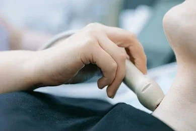
A hand-held ultrasound transducer is smoothly glided along the carotid artery pathway.
The exam is completely non-invasive, pain-free, involves no radiation or contrast dyes, and typically takes 30 minutes.
Pre-Appointment Reminder / Next Steps
Have Questions About Your Upcoming Test?
If you have specific questions about how to prepare or what to wear for your neurodiagnostic appointment, please contact the Florida Neurovascular Institute office at (813) 250-9101.